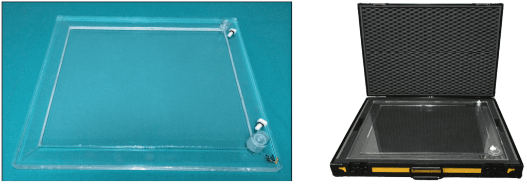
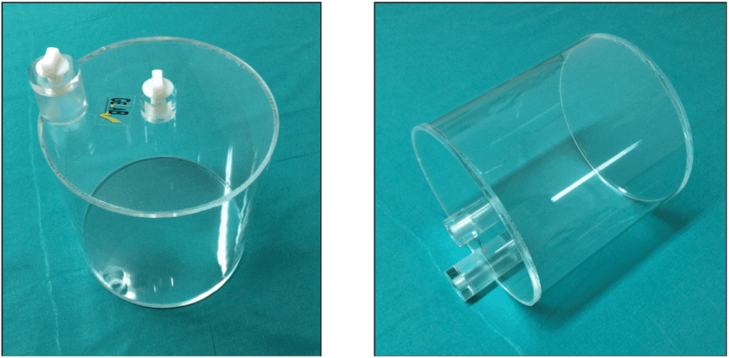
The flood phantom provides a simple and efficient means of obtaining an optimal assessment of the camera with respect to the uniformity of the response across the entire surface. Its rectangular design contains a sealed central cavity, into which a radioactive solution is introduced through an injection port and homogenized by agitation. Once the activity is evenly distributed, the filling process is completed, and the air remaining inside is directed toward the filling port, resulting in a uniform source with a bubble-free field of view.
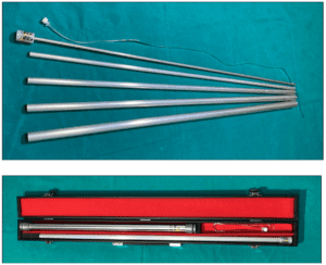
The phantom can be supplied with a “flight case” type transport container.
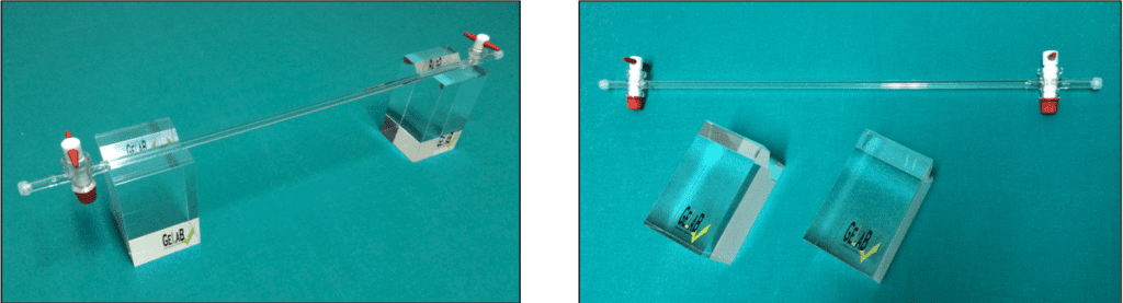
Under clinical conditions, the dead time of a scintillation camera is a function of radiation scatter from the source. The Adams phantom simulates the in vivo scatter of gamma radiation from Tc-99m, producing a typical spectrum similar to that observed in an “in vivo” myocardial test.
This phantom is used for the determination of dead time and temporal resolution of gamma cameras.
The pixel size is determined based on the acquisition matrices used, both along the “X” and “Y” axes, which is highly relevant in the attenuation correction calculation algorithm. It is a fundamental test in the quality control of SPECT.
The phantom is capable of obtaining measurements along both axes in a single acquisition, optimizing the machine’s operating time.
![]()
The sensitivity of a gamma camera is a characteristic parameter that expresses, for a certain time interval, the counts detected per unit of activity.
The sensitivity phantom is designed to obtain counting data with low activity and source thickness, being very easy to handle and minimizing the possibility of contamination caused by the use of Petri dishes.
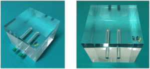
Tomography (PET/SPECT) imaging systems require periodic uniformity testing.
For the determination of tomographic uniformity, the phantom must be filled with a homogeneous solution of water and Tc-99m, which is achieved using a two-hole filling system with bubble trapping.
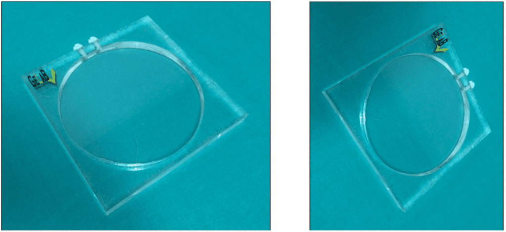
The image obtained from a linear source with a diameter of 1 mm, and its processing through count profiles, provides FWHM (Full Width at Half Maximum) values used to assess this test. This determination is performed along both the “X” and “Y” axes.
Extrinsic spatial resolution is strongly dependent on the distance from the linear source to the detector system. The phantom is equipped with supports that maintain this distance constant across successive measurements.
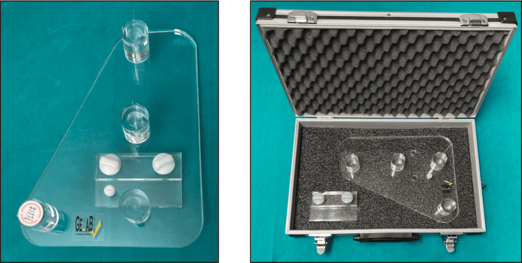
The set of phantoms for gamma camera quality control can be supplied with a “flight case” type transport container.
![]()
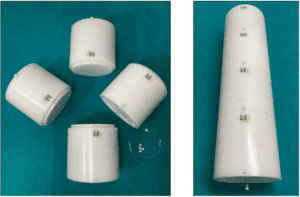
Made of a solid cylinder with a diameter of 20 cm and a length of 70 cm, constructed from HDPE, featuring a 6.5 mm diameter hole parallel to the central axis of the cylinder, displaced 4.5 cm from the axis, for inserting the refillable linear source. It is composed of four detachable sections to facilitate transportation/storage.
This phantom is used for determining the scatter fraction, loss fraction, and random events. It measures the accuracy of random event corrections and event losses. It also measures dead time and random events generated at different activity levels, according to NEMA standards.
The spatial resolution of a system defines its ability to distinguish two point sources as separate in a reconstructed image. It is defined as the FWHM (Full Width at Half Maximum) of a point spread function (PSF) and is calculated from the line profile through a reconstructed image of a point source.
This device consists of a PMMA support with a positioning system, suitable for: glass capillary tubes, MMS05-type sources (1″ x 0.25″ disc), and MMS09-type sources (10 mm cube). It allows for the placement of all three point sources in either of the two sets of positions described in the NEMA NU 2-2007 and NEMA NU 2-2012 standards.
Sensitivity is the number of counts per unit of time and activity. The measurement of sensitivity should be independent of factors such as scatter, attenuation, count losses, and random events.
This device consists of five concentric aluminum tubes, each 70 cm in length with a wall thickness of 1.22 mm, a refillable tube made of transparent plastic, 80 cm in length with an inner diameter of 1 mm and an outer diameter of 3 mm, and an extendable aluminum tube for placement along the FOV axis, in accordance with the NEMA standards.
The spatial resolution of the probe is an essential parameter for distinguishing nearby radiation sources, such as the injection site and the sentinel lymph node.
Angular resolution characterizes the dependence on the angle at which the probe is oriented relative to the source.
Lateral resolution quantifies the probe’s ability to detect two very close sources as distinct sources, rather than as a single extended source.
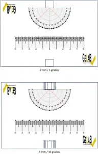
This template can be printed on adhesive paper, cut out, and affixed to a lightweight matrix (e.g., expanded polystyrene) where holes or drills can be made at the indicated points.
This creates a support and positioning device for the probe, fixed on one side for spatial resolution and on the other side for angular resolution. This setup is designed for the measurement of a point source (at a distance of 30 mm), which must be placed at each of the indicated points.
- For lateral resolution: up to x = ± 20 mm, in 2 mm intervals, and from x = ± 20 mm to x = ± 50 mm, in 5 mm intervals (or simplified to 5 mm intervals).
- For angular resolution: up to α = ± 30°, in 5° intervals, and from α = ± 30° to α = ± 90°, in 10° intervals (or simplified to 10° intervals).
Click on the image to download the template